


- New fingerprint breakthrough by forensic scientists
June 2, 2008 09:30 PM
Forensic scientists at the University of Leicester, working with Northamptonshire Police, have announced a major breakthrough in crime detection which could lead to hundreds of cold cases being reopened.
The University's Forensic Research Centre has been working with Northamptonshire Police's scientific support unit to develop new ways of taking fingerprints from a crime scene.
Researchers in the University Department of Chemistry and the Police's scientific support unit have developed the method that enables scientists to 'visualise fingerprints' even after the print itself has been removed. They conducted a study into the way fingerprints can corrode metal surfaces. The technique can enhance – after firing– a fingerprint that has been deposited on a small calibre metal cartridge case before it is fired.
"Wiping it down, washing it in hot soapy water makes no difference - and the heat of the shot helps the process we use.
"The procedure works by applying an electric charge to a metal - say a gun or bullet - which has been coated in a fine conducting powder, similar to that used in photocopiers.
"Even if the fingerprint has been washed off, it leaves a slight corrosion on the metal and this attracts the powder when the charge is applied, so showing up a residual fingerprint...
- Cartilage regeneration
Date: 05/06/2008
Bioengineers at Rice University have discovered that intense pressure -- similar to what someone would experience more than a half-mile beneath the ocean's surface -- stimulates cartilage cells to grow new tissue with nearly all of the properties of natural cartilage. The new method, which requires no stem cells, may eventually provide relief for thousands of arthritis sufferers."This tissue-engineering method holds promise not only for cartilage but also for tissues to repair bladders, blood vessels, kidneys, heart valves, bones and more," said lead researcher Kyriacos Athanasiou, Rice's Karl F. Hasselmann Professor of Bioengineering.
The findings appear this week in the journal PLoS ONE. They are the latest from the emerging field of tissue engineering, a new discipline that aims to capitalize on the body's innate healing abilities to develop new ways of growing tissues that can be used to surgically repair wounds without risk of rejection.
Cartilage, a tissue in the human body that cannot heal itself, has long been a target of tissue engineers. Cartilage is the skeleton's shock absorber, and its stiffness, strength and other mechanical properties derive not from living cartilage cells but from the densely woven matrix of collagen and proteoglycan that surrounds them. This extracellular matrix, or ECM, is produced during cartilage development in children, but cannot be repaired following injury in adulthood.
Injured cartilage often serves as the focal point for arthritis formation, so tissue engineers have long sought a means of growing new cartilage that can be transplanted into adults to repair damaged joints before arthritis can develop. Unfortunately, cartilage is difficult to engineer, in part because there are no natural healing processes to mimic.
Athanasiou's Musculoskeletal Bioengineering Laboratory has focused on cartilage for more than 10 years, and he said the new process is the first he has studied that produces cartilage that's almost identical to the body's own tissue.
"The combination of hydrostatic pressure and growth factors used in this process result in an engineered cartilage ECM with properties nearly identical to that of native cartilage," he said. "This research appears very promising for treating arthritis, as cartilage can now be produced in our lab that is almost identical in composition to native tissue."
So far, the process has been tried only with cells from cows and has yet to be tested in live animals. Athanasiou cautions that it will be several years before the process will be ready for clinical testing in humans.
The new findings are based on three years of data collected by graduate student Benjamin Elder, who is simultaneously earning a doctorate in bioengineering at Rice and a medical degree at Baylor College of Medicine under Rice and Baylor's Medical Scientist Training Program...
- Hairy blobs in acidic hell
Date: 05/06/2008
Close-up, they look like something out of a 1950s B-movie. Colonies of fossilised creatures, dubbed "hairy blobs", have been discovered in one of the harshest environments on Earth. The find may turn out to be crucial for spotting signs of extraterrestrial life in rocks on other planets.Kathleen Benison, a geologist at Central Michigan University, Mount Pleasant, led a team that studied the sediments formed by acidic and very salty lakes in modern day Western Australia, and those deposited around 250 million years ago in North Dakota. It is very difficult to survive in such a tough environments and few signs of life have ever been found in these sorts of lakes.
Inside the halite and gypsum "evaporate" minerals, which form as the lake waters dry up, Benison and colleagues found previously unknown fossilised blobs at both the modern and ancient sites, ranging in size from 0.05 to 1.5 millimetres. They were made up of a mix of inorganic crystals and "hairs" stuck together in a mass (pictured). They named them hairy blobs.
The team argues that each hair was in fact a separate microorganism because the hair fossils are made of disordered graphite which, unlike inorganic graphite, has irregular layers that suggest it was once a live organism..
Many of the hairs are coated with crystals of gypsum, a calcium sulphate mineral. This link with gypsum suggests that the microorganisms were fuelled by chemical interactions with sulphur in the acidic water - which helped the gypsum to form.
The team also found previously undescribed microorganisms in the lake water, which they say may be the cells that end up as fossilised hairs (Astrobiology, DOI: 10.1089/ast.2006.0034).
Conditions in acidic saline lakes such as those studied by the team are thought to be similar to those on ancient Mars. The many probes currently exploring the Red Planet have discovered that Martian seas and lakes, such as those once found at Meridiani Planum, were strikingly similar in terms of acidity, salinity and the minerals and sediments present.
Benison says the hairy blobs from the Permian halite seem well preserved. "This argues for long-term preservation of microfossils in halite elsewhere, perhaps even on Mars." Had the organisms lived on Mars, she says, the inorganic minerals surrounding them would have acted as protection from the ultraviolet radiation there...
- Talking to cells
June 5, 2008 08:17 PM
For an organism to develop and function, the individual cells must exchange information, or communicate, with each other. Is it possible to learn their language and "talk to" the cells?
Yes it is: Cameron Alexander and George Pasparakis at the University of Nottingham (UK) have been able to facilitate a conversation between bacterial cells and artificial polymer vesicles. In the journal Angewandte Chemie they report that this first communication occurred by way of sugar groups on the vesicle surface. The vesicles subsequently transfer information to the cells—in the form of dye molecules.
Complex structures made of many sugar components on the surfaces of cells are the "language" used for processes such as cell recognition, for example, in the differentiation of tissues or the identification of endogenous cells and foreign invaders. Scientists would like to be able to use this glycocode to "address" target cells and to intervene directly in cellular processes to treat diseases or to guide regeneration of damaged tissues.
The British scientists took an interesting route to learn more about the "language" of cells: they constructed vesicles, tiny capsules whose outer shell is made of special polymer building blocks. Their special trick: the polymer chains are equipped with side chains bearing glucose units that wind up being exposed on the vesicle surface.
The researchers brought the vesicles together with bacteria that have glucose-binding proteins on their surface. The behavior of the bacteria varies depending on the polymer's composition and the size of the vesicles. Among the bacteria were some individuals that enter into very strong bonds with large vesicles. These associated bacteria are then in a position to receive molecular "information" from the vesicles: dye molecules that were previously placed in the vesicles transferred specifically into the interior of these bacteria...

The British scientists took an interesting route to learn more about the "language" of cells: they constructed vesicles, tiny capsules whose outer shell is made of special polymer building blocks. Their special trick: the polymer chains are equipped with side chains bearing glucose units that wind up being exposed on the vesicle surface. (Credit: Copyright Wiley-VCH)
Full Article
- Measuring the footprint of cells
June 7, 2008 04:22 PM
Even the slightest differences are important in competitive sport: To improve a ski jumper's performance, the trainer can analyze the jump very accurately using force sensors. Researchers in Jena and Bremen are planning something similar. However, their work is not with athletes but with tiny somatic cells. The experts have developed a low-cost optical sensor to measure the force with which migrating cells push themselves away from an underlying surface. Force analysis devices like these could one day help to identify specific cell types – more reliably than using a microscope or other conventional methods.
The sensor is the outcome of an EU project. It consists of a smooth surface that is studded with 250,000 tiny plastic columns measuring only five microns in diameter, rather like a fakir's bed of nails. These columns are made of elastic polyurethane plastic. When a cell glides across them, it bends them very slightly sideways. This deflection is registered by a digital camera and analyzed by a special software program. The researchers working with project manager Dr. Norbert Danz of the Fraunhofer Institute for Applied Optics and Precision Engineering IOF in Jena have already shown that their 'Cellforce' sensor works. It will be the task of initial biological tests to show how different cell types behave. "Analysis of cell locomotion is important for numerous applications," says Danz. "It could be used to check whether bone cells are successfully populating an implant, or how well a wound is healing."
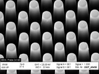
The sensor is covered with 250,000 tiny plastic columns only five microns in diameter. When a cell creeps across the tips of the columns, it presses each column very slightly sideways. Credit: © Fraunhofer IFAM
- Microspheres to carry hydrogen, deliver drugs, filter gases and detect nuclear development
June 7, 2008 04:22 PM
What looks like a fertilized egg, flows like water, gets stuffed with catalysts and exotic nanostructures and may have the potential of making the current retail gasoline infrastructure compatible with hydrogen-based vehicles of the future – not to mention also contributing to arenas such as nuclear proliferation and global warming?
The answer is contained in the June issue of The Bulletin, the monthly magazine of The American Ceramic Society, which carries the first news of a never-before-seen class of materials and technology developed by scientists at the Savannah River National Laboratory.
The full article can be downloaded at
This unique material, dubbed Porous Wall-Hollow Glass Microspheres (PW-HGM), consists of porous glass 'microballoons' that are smaller than the diameter of a human hair. The key characteristic of these 2-100 micron spheres is an interconnected porosity in their thin outer walls that can be produced and varied on a scale of 100 to 3,000 Angstroms.
SRNL Researchers G.G. Wicks, L.K. Heung, and R.F. Schumacher have been able to use these open channels to fill the microballons with gas absorbents and other materials. Hydrogen or other reactive gases can then enter the microspheres through the pores, creating a relatively safe, contained, solid-state storage system...
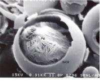
SRNL researchers removed the top of a glass microsphere to show how palladium has easily passed through the sphere's pores and assembled itself into a new nanostructure.
- Molecular 'Ratcheting' Of Single Ribosome Molecules Observed In Act Of Building Proteins
ScienceDaily (Jun. 6, 2008)
Researchers have reported that they are the first to observe the dynamic, ratchet-like movements of single ribosomal molecules in the act of building proteins from genetic blueprints.
Their study, published in the journal Molecular Cell, reveals a key mechanism in the interplay of molecules that allows cells to build the proteins needed to sustain life.
Cells use a variety of tools to build proteins, beginning with messenger RNA, a ribbon-like molecule that codes for the sequence of amino acids in the protein. Another molecule, transfer RNA (tRNA) is uniquely qualified to read this code, but can do so only within the confines of the ribosome. Transfer RNAs bring individual amino acids into the ribosome where they are assembled into proteins. Various other proteins also participate in the process.
When protein translation occurs, single tRNAs enter specific sites in the ribosome, read the code and deliver their amino acids – one by one – to a growing protein chain. The ribsome transits along the messenger RNA as the protein is built, releasing the “deacylated” tRNA through an exit site.
A ribosome is made up of two subunits composed of ribonucleic acids (RNAs) and about 50 individual proteins....
- New Way To Think About Earth's First Cells
ScienceDaily (Jun. 6, 2008)
A team of researchers at Harvard University have modeled in the laboratory a primitive cell, or protocell, that is capable of building, copying and containing DNA.
Since there are no physical records of what the first primitive cells on Earth looked like, or how they grew and divided, the research team's protocell project offers a useful way to learn about how Earth's earliest cells may have interacted with their environment approximately 3.5 billion years ago.
The protocell's fatty acid membrane allows chemical compounds, including the building blocks of DNA, to enter into the cell without the assistance of the protein channels and pumps required by today's highly developed cell membranes. Also unlike modern cells, the protocell does not use enzymes for copying its DNA.
Led by Jack W. Szostak of the Harvard Medical School, the research team published its findings in the June 4, 2008, edition of the journal Nature's advance online publication.
"Szostak's group took a creative approach to this research challenge and made a significant contribution to our understanding of small molecule transport through membranes," said Luis Echegoyen, director of the NSF Division of Chemistry.
Some scientists have proposed that ancient hydrothermal vents may have been sites where prebiotic molecules--molecules made before the origin of life, such as fatty acids and amino acids--were formed.
When fatty acids are in an aqueous environment, they spontaneously arrange so that their hydrophilic, or water-loving, "heads" interact with the surrounding water molecules and their hydrophobic, or water-fearing, "tails" are shielded from the water, resulting in the formation of tiny spheres of fatty acids called micelles.
Depending upon chemical concentrations and the pH of their environment, micelles can convert into layered membrane sheets or enclosed vesicles. Researchers commonly use vesicles to model the cellular membranes of protocells...

Above is a three-dimensional view of a model protocell approximately 100 nanometers in diameter. The protocell's fatty acid membrane allows nutrients and DNA building blocks to enter the cell and participate in non-enzymatic copying of the cell's DNA. The newly formed strands of DNA remain in the protocell. (Credit: Janet Iwasa, Szostak Laboratory, Harvard Medical School and Massachusetts General Hospital)
- Possible New Approach To Purifying Drinking Water, Thanks To Genetic Tool
ScienceDaily (Jun. 9, 2008)
A genetic tool used by medical researchers may also be used in a novel approach to remove harmful microbes and viruses from drinking water.
In a series of proof-of-concept experiments, Duke University engineers demonstrated that short strands of genetic material could successfully target a matching portion of a gene in a common fungus found in water and make it stop working. If this new approach can be perfected, the researchers believe that it could serve as the basis for a device to help solve the problem of safe drinking water in Third World countries without water treatment facilities.
The relatively new technology, known as RNA interference (RNAi), makes use of short snippets of genetic material that match -- like a lock and key -- a corresponding segment of a gene in the target. When these snippets enter a cell and attach to the corresponding segment, they can inhibit or block the action of the target gene. This approach is increasingly being used as a tool in biomedical research, but has not previously been applied to environmental issues.
"Pathogens, whether bacterial or viral, represent one of the major threats to drinking water in developed and undeveloped countries," said Sara Morey, a Ph.D. candidate in the lab of Claudia Gunsch, assistant professor of civil engineering at Duke's Pratt School of Engineering. "Our data showed that we could silence the action of a specific gene in a fungus in water, leading us to believe that RNAi shows promise as a gene-silencing tool for controlling the proliferation of waterborne bacteria and viruses."
Morey presented the results of her experiments June 3, 2008, during the annual meeting of the American Society of Microbiology in Boston...


No comments:
Post a Comment